Fluorescence Microscope
Prototypes
Fluorescence microscopy is a technique used to analyze biological structures in a sample using a white lamp, and either organic or inorganic fluorophores such as dyes to excite a photo-emissive reaction, which is observed using an optical bandpass filter and a dichroic mirror. Various biochemical industries rely on fluorescence microscopy for the performance of molecular imaging to support medical diagnostics or research into cellular structures and nanomolecular activity.
Delta Optical Thin Film has previously discussed the principles of, and hardware implemented in modern fluorescence microscopy, but this article will explore some of its immediate applications and implications of further study in the field in more detail..]]
Version 01 - First Prototype by Urs / GaudiLabs
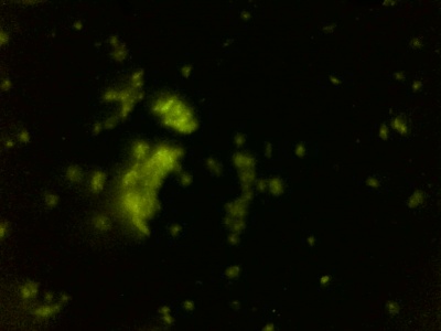
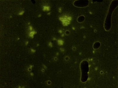
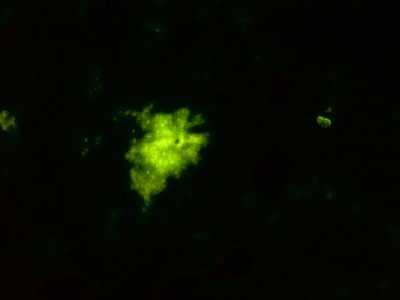
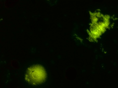
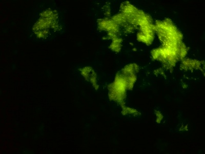
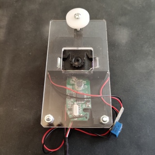
The first prototype used both some parts 3d printed, and others with the laser cutter. The camera module was not properly enclosed. First results really promising.
GlowScope (Version 02) - redesigned by Urs and tested and assembled by pin
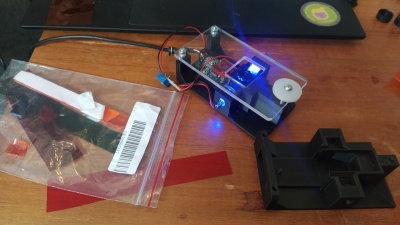
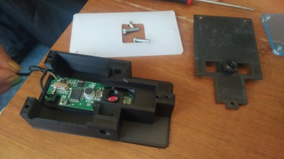
To improve the design, a full matt black enclosed was designed and 3d printed to properly host the camera module and 2 LED from both sides. A slider inside the enclosure allows to change filters.
Parts needed:
Logitech C270 Webcam, 2x Blue LED 5mm, 1 x 200-1000 Ohm resistor, some wire, Blue and orange filter foil, M3 screws, 4x Rubber feets
All the files and material lists are on github: https://github.com/GaudiLabs/GlowScope
Version 03
After experiences with Version 02, there is still room for improved designs.
Following ideas:
- 2 different LEDs. how does one-sided illumination work?
- improve filter slider (what if it "clicks" into place?)
- Make more tests with properly prepared stained and fixed samples.
This electronically switchable filter module from ali looks promising!
DIY Fluorescence microscopy
Introduction
Uses
Fluorescence Microscopy for Cell Labeling
Labeling cell structures with fluorophores such as fluorescent dyes drastically increase the observable properties of biological or chemical structures. These can be modified or conjugated to exhibit specific targetability for use in various medical applications. Fluorophores with high specificity are routinely used in medical diagnostics, with increasing research finding further applications for fluorescent cell labeling. For example: researchers have successfully used fluorescence microscopy to track and analyze viral DNA in cells at a local level and further connect that to cellular anti-viral defenses. This has potentially tremendous implications for future viral analysis and categorization.
Fluorescence Microscopy for Protein Characterization Intra-cellular dynamic protein interactions are crucial for a range of critical biological processes, acting as catalysts for metabolic reactions or responding to stimuli. Protein interactions are typically observable using dedicated instrumentation capable of analyzing the samples at the nanometer scale. However, transient interactions of proteins display weaker signals which are typically transparent, even to fluorescence microscopy instrumentation. The implementation of fluorescent protein biosensors is a novel approach to observing weaker protein interactions, which has found broad applications in varying fields of research. Researchers have utilized fluorescent proteins to observe intra-cellular reactions in the hopes of gaining insight into various transmission reactions, by analyzing the specific wavelengths emitted by excited cells during protein synthesis. Such studies have tremendous potential to provide insights into how irregular translations in a cell can result in developmental problems and disease transmission – yet they also require exceptionally sensitive instrumentation and highly tunable chromatic filters to accurately assess data.
fluorescence microscopy applied to Biology
      • Within microscopy
      • material imaging
      • medical imaging
      • electron microscopy (transmission or scanning)
      • scanning probe microscopy
      • light microscopy
      • bright field microscopy
      • fluorescence microscopy
      • wide field microscopy
      • con focal microscopy
Light is an electromagnetic wave created by photons
      • A point charge at rest produces an electric field
      • A point charge moving with constant speed produces an electric field and a magnetic field
      • A point charge moving with a varying speed (acceleration) produces an electromagnetic wave which can travel at 3.108m/s though empty space
      • Light is an electromagnetic wave created by accelerating photons
      • How the speed of the charge changes determines the length of the wave, how energetic it is and how focused the beam can be
